Whether you're going for your very first ultrasound or you have been to several in the past, learning common ultrasound terminology can be quite helpful. You'll better understand what the doctors are talking about, and it will help you ask questions to further your knowledge about your medical situation. Keep reading for examples of ultrasound terminology that can help you in your next doctor’s visit.
Common Ultrasound Terminology
There is an abundance of ultrasound and medical terminology used during the ultrasound process. Learn the definitions for some of the keywords and phrases.
- A-mode (or amplitude modulation) - an imaging technique that focuses on the different heights of amplitude spikes (such as in a heartbeat monitor)
- acoustic enhancement (or posterior enhancement) - the ability for sound waves to travel without return echoes because an anechoic space is full of fluid (such as cysts or the bladder), creating a black space in the sonogram
- acoustic shadowing - area in which sound waves encounter a dense structure, such as a tumor or fetus, and all waves are reflected
- aliasing phenomenon - imaging error that occurs in color flow Doppler and pulsed wave Doppler scans; sampling rate is insufficient to record direction and velocity
- anechoic structure - body chamber without echoes; fluid-filled structures that appear black on the sonogram
- anterior (or ventral) position - in front
- attenuation - the tendency for ultrasound waves to decrease as they travel through tissues
- axial (or longitudinal) resolution - minimum distance between two reflectors along the ultrasound beam direction
- B-mode (or brightness modulation) - an imaging technique that focuses on the brightness of the echo, not the amplitude spikes (such as in a typical 2D ultrasound)
- color flow Doppler - Doppler scan that measures blood flow by attributing color to different velocities of movement
- coronal scanning plane - the ultrasound beam enters the body from the lateral (right or left) direction
- distal position - away from the point of origin or trunk
- Doppler shift/Doppler effect - change in frequency as the transducer moves away from the source of the sound echoes
- Doppler ultrasound - procedure named after physicist Christan Doppler in which sound waves bounce off circulating red cells to measure blood flow; often used to detect the heartbeat of a fetus
- gel couplant - gel used in an ultrasound scan; functions as the medium through which sound energy is transmitted between the transducer and the patient's skin
- hyperechoic - structures that return greater echoes of sound waves and appear bright white on a sonogram
- hypoechoic - structures that return weaker echoes of sound waves and appear gray or black on a sonogram
- inferior position - lower, away from the head
- lateral position - toward the side
- lateral resolution - minimum distance between two reflectors perpendicular to the ultrasound beam direction
- M-mode (or motion mode) - early ultrasound modality that shows the time-motion display of the ultrasound wave; displayed as a single scan line, not a visual image
- medial position - toward the middle or center
- mechanical probes - a transducer that has a motor inside
- posterior (or dorsal) position - toward the back
- power Doppler - highly sensitive Doppler technique that detects moving matter
- proximal position - toward the point of origin or trunk
- pulsed-wave Doppler - Doppler technique that uses short and quick pulses of sound to measure the velocity of blood in a specific location
- reverberation artifact - event in which sound waves become trapped between two parallel structures and bounce between them before returning as echoes
- sagittal scanning plane - the ultrasound beam enters the body from anterior or posterior (front or back) position
- scanning plane - direction at which the ultrasound beam enters the body
- sonogram - image produced during an ultrasound
- sound waves - waves of energy that are transmitted from a transducer through the gel couplant, skin and tissue; they return as echoes after being reflected by structures in the body
- speckle noise - noise interference that reduces the quality of the image resolution in a sonogram
- superior position - higher, toward the head
- time gain compensation (TGC) - a technique in which signal gain increases as time passes (used to overcome attenuation)
- transducers - devices that produce sound waves in ultrasounds and send return echoes to a computer, which compiles them into a sonogram
- transverse scanning plane - the ultrasound beam enters the body from anterior, posterior or lateral (front, back, right, or left) position; scans in a diagonal or transverse angle
- ultrasound frequency - "ultra" or inaudible sound frequency at which ultrasounds operate (ranges from 2.5 megahertz to 15 megahertz; humans can hear sounds no higher than 20,000 hertz)
- ultrasound scans - a medical procedure in which sound waves are beamed into the body, and their return echoes enable technicians to visualize what is happening under the skin
- velocity of ultrasound - the speed at which sound waves can travel through different parts of the body, measured in meters per second (1540 m/sec for soft tissue, 1580 m/sec, 4080 m/sec for bone)
Ultrasound vs. Sonogram
You may hear the terms ultrasound and sonogram used interchangeably when discussing an ultrasound scan. However, they are two parts of the same procedure. The ultrasound is the procedure itself, while the sonogram is the picture on the screen produced by the ultrasound. Think of it as process (ultrasound) vs. results (sonogram).
Types of Ultrasounds
Depending on what your doctor needs to see, they may recommend a variety of ultrasound procedures. Some of these procedures include:
- 2D ultrasound - ultrasound scan that creates a two-dimensional cross-sectional (flat) image
- 3D ultrasound - ultrasound scan that uses angles to create a three-dimensional (round) image
- 4D ultrasound - ultrasound scan that uses angles to resemble movement under the skin in a four-dimensional (moving) image
- aortic ultrasound - scan of the aorta, which is the body's main blood vessel that carries blood away from the heart; typically used to rule out an abdominal aortic aneurysm
- cranial ultrasound - scan of the brain; typically used on babies before their fontanelles (soft spots) close
- echocardiogram - ultrasound of the heart and cardiac blood vessels; used to measure blood flow through the heart and how well it functions
- endoscopic ultrasound (or "echo") - ultrasound procedure in which a transducer goes down the esophagus to measure the respiratory and digestive systems; typically used to diagnose diseases or disorders of the lungs, stomach, lymph nodes, upper digestive tract, liver, and pancreas
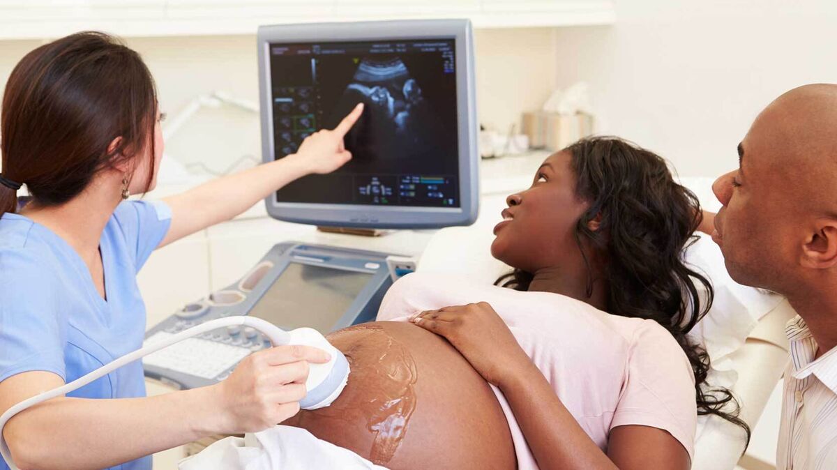
- obstetric (OB) ultrasound - scan of the lower abdomen to detect and monitor a growing fetus during pregnancy; typically occurs during 12-week scans (fetal size and development) and 20-week scans (fetal organs and development)
- pelvic ultrasound - scan of the pelvic area to monitor conditions of the female reproductive system (different from the transvaginal ultrasound, which uses a vaginal probe)
- renal ultrasound - ultrasound of the kidneys; used to determine the presence and function of the kidneys as well as the presence of kidney stones, tumors or cysts
- thyroid ultrasound - scan of the thyroid gland in the neck; typically used to detect cysts, nodules or tumors, or to determine whether the thyroid is overactive or underactive
- transabdominal ultrasound - scan of the abdominal cavity, which includes the pancreas, kidneys, bladder, spleen, and gallbladder
- transcranial Doppler - Doppler ultrasound that measures the velocity of blood flow in the brain; used to test for brain aneurysms, stroke risk and blood vessel blockage (intracranial stenosis)
- transrectal ultrasound - scan of the rectal area and prostate by a rectal transducer probe
- transvaginal ultrasound - ultrasound scan in which a vaginal probe bounces sound waves through the pelvis to examine organs in the female reproductive system; typically used in early pregnancy, ectopic pregnancy, ovarian cysts, IUD checks, or other gynecological concerns (different from a pelvic exam, which uses an exterior transducer)
What Ultrasounds Can Scan For
Performing an ultrasound is a good first step before having an MRI (Magnetic Resonance Imaging), CT scan (Computerized Tomography) or more invasive procedure. Technicians use ultrasounds to scan for the following conditions:
- abdominal pain
- abscess in the body
- aneurysm
- appendicitis
- atrial fibrillation
- blood clot
- bowel obstruction
- breast lumps/legions
- congestive heart failure
- cyst
- deep vein thrombosis (DVT)
- ectopic pregnancy
- endometriosis
- enlarged organs
- fatty liver disease
- fetal abnormalities
- fibroids
- gallstones
- hypertension
- infection
- placenta previa
- pregnancy
- stenosis
- testicular torsion
- thrombosis
- tumors
Know More About Ultrasounds
Getting an ultrasound is a generally safe form of preventative care. Now that you know more about the terminology associated with ultrasounds, learn all about the abbreviations used in pregnancy ultrasounds. Or, if you're curious about different medical fields, take a look at a helpful medical abbreviations list.